Japanese Page
Iguanas will not talk to you, will not complain about the food you are giving, will not tell you that she has a too-too tummy.
Fecal examination
You have to watch them carefully and should not miss any information. One of the best information we can get is from their poop. If you love your animal, love her poop too. Poop is the only message given from them daily.
If you find their droppings, take them our carefully and observe. It will be convinient if you prepare tissue box and a disposable toothpick near by.
Naked Eye Observation
First check the quantity. Does it seem normal compare to the otherday's poop? If their poop is obviously thin, thick, watery, dried, slimy, stinky, or abnormal color than usual, it might be a signal of some kind of disease.
Watch them daily and be aware of the change.Next, get your eyes close to their dropping and scan very carefully. Nematodes can be found by naked eye. They are 5 to 10mm in length but very thin like a small piece of thread. There are many other fibrous matter from their food that may look similar to namatodes but if you look closely or if you can pick them up by the toothpick and place it on black paper, you can see that namatodes moves but vegetable fibers don't. Usulally, their movement are very slow so you may have to watch them for over a minute.
If you find something like this, consult your veterinarian. If it is difficult to take your iguana, bring the fresh poop to the vet. Film case will be a good container.
If you don't have a vet who can treat reptiles near by, it is possible to check it yourself. You will need a microscope but you don't have to buy a super magnification set like 600x,1000x etc.. I will recommend you to buy a system microscope so you can start by least set and if you are interested, you can upgrade later. At the beginning, you will just need the body and 2 objective lenses and an eye-piece.
Select 10x and 40x oblective lenses and 10x eye-piece. This will make total magnification of 100x and 400x. 10x objective lens is used to search for nematodes and nematode eggs. 40x objective lens is used to observe protozoa.
There are many kinds of parasites. First they are devided into groups of external parasites and internal parasites.
External parasites includes mites and tics.
Internal parasites are devided into further groups. Some lives in digestive tract, some lives in organs, and some lives in cells. Here we will discuss about the parasites which only lives in digestive tract. For further study, I recommend you to read books about reptile parasites.
Internal Parasite
Parasites which are tend to live in iguanas usually not so much pathogenic. Iguanas which are untreated by parasiticide mostly carry many kinds of parasites. If your iguana has some parasites, that does not mean that your iguana will die in a day or a week. It usually does nothing if your iguana is healthy. But once your iguana got sick by other cause and if it's immunity gets down, carrying a parasite may be pathogenic. So, you don't need to rush but I recommend you to treat them when they are healthy.
You can check an find their parasites but be sure not to treat them by yourself. Do not use cat's or dog's parasiticide by guessing the dosage. Consult your veterinarian and treat them by prescribed medicine.
After the fecal examination, wash your hands throughly by running water and some antibiotic soap.
Protozoan
In a watery feces or diarrhea excretion, you may find many kind of protozoa. Most common protozoan found in iguana feces is trichomonads sp. Metronidazole(Fragyl) is a common parasiticides to treat trichomonads but I personally do not think it is necessary to treat them. It is difficult to treat them completely and if you try to kill trichomonads, that means you also have the possibility of killing all the beneficial bacteria for their digestion system. Also the pathogenicity of trichomonads are unknown. Further research is necessary but they might have a symbiotic relationship with iguanas.
We are keeping an 8 years old iguana and trichomonads are found in her feces since she was a juvenile. I gave up to treat her trichomonads in a first few month from above reason and she is still carrying bunch of them. She is healthy and can't find any pathogenic symptoms. I should not conclude yet but maybe trichomonads are not parasites and helping their digestion intead.
I will recommend you to consult your veterinarian.
Nematode
If you find a thread looking very thin worm in iguana's feces, place it on a slide glass and watch them by low magnification lens. If you find a worm like the picture right, it is a nematode.
If you can't find the nematode itself, existance of nematode can be convinced by finding their eggs in iguana's feces. Eggs of nematodes are about 140 micro-meter by 80 micro-meter in an ellipsoidal shape. You can only find them under the microscope but you will find it easy to find because they lay so many eggs everyday. If you find any of these, consult your veterinarian.Wild caught iguanas usuall carry nematodes and it is thought to be not so pathogenic in usual case, but once the host gets weak by some other reason and force of immune system gets low, they will be pathogenic. They will be harmful to their digestive system. So treat them when the host is healthy.
Nematodes usually does not need an intermediate host so they can reproduce in the host and also be able to infect other iguanas by ingesting nematode eggs orally.If you have more than one iguanas and if you find nematode or nematode eggs, you will have to treat all possible hosts.
Now a days, treating nematodes are quite simple. You can get rid of them completely by 2,3 weeks.Cestode
I have heard of parasitic cestodes hosting an iguana but I have not seen any by myself. I guess cestode in iguana is a very rare case.
Since the movement of protozoa are rapid, it is better to go through the fixation and staining process. Most protozoa are invisible and difficult to see the details under the microscope. By good staining process, you will be able to see the nuclear and count the flagella of protozoa. You can make a permanent prepared specimen which will be accademically valuable.
We will use trichomonads for an example.
Now you will have a parmanent specimen. Look through it under your microscope. You will find a new universe in a 18mm by 18mm square.
- Dissolve small piece of feces in a Ringer's solution.
- Take 1 drop of this solution and make a thin layer as possible on new slide glass.
- Quickly dry the expanded solution. You can use the cool air dryer.
- Soak the slide glass in 100% methyl alcohol for few minutes.(fixation)
- Stain. I usually use methyl alcohol diluted Giemsa stain.
- Dehydrate. Prepare 50%, 70%, 100% ethyl alcohol. Soak the slide glass from low percentage solution to pure alcohol. It is better to make 2 100% container and soak it twice by using both container.
- Soak the slide glass in 100% xylol.
- Dry and cover it by cover glass using balsam.
- Leave the slide glass on the leveled box for a week or more until balsam gets hard.
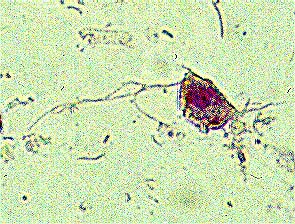
Trichomonas
Dissolved in 0.8% Ringer's solution
Smear on slide glass
Dried by cool air
Methyl alcohol fixation
Stained by Giemsa's stain
Dehydrated by ethyl alcohol, xylol
Sealed with balsam
Objective lens:OLYMPUS PCD-Ach 40x
Photographic lens:OLYMPUS LD 3.3x
Camera:PENTAX 645 body, self-made adaptor
Film:FUJI VELVIA 120
Trichomonad has an ovary pear shaped body. They have 3 to 5 flagella. When they are alive, you will find it difficult to count their flagella because each have rapid movements. But after fixation it is easily countable. This specimen is Tri-trichomonas which has 3 fragella. Undulating membrane can be seen by better fixation and staining.
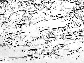
Spermatozoon
Direct smear on slide glass
Dried by cool air
Methyl alcohol fixation
Stained by Giemsa's stain
Dehydrated by ethyl alcohol, xylol
Sealed with balsam
Objective lens:OLYMPUS DPLAN 10x
Photographic lens:OLYMPUS LD 5x
Camera:PENTAX 645 body, self-made adaptor
Film:FUJI VELVIA 120
B/W image process
The shape of spermatozoon of iguanas are different from the one from humans. Head part is long in a shape like stringbeans. If you have a male iguana, you will have a chance to watch the live active spermatozoons dynamically. They can be easily collected in the mating season without any harm.
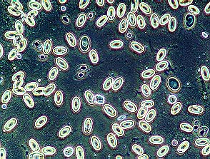
Erythrocyte
Direct smear on slide glass
Dried by cool air
Methyl alcohol fixation
Stained by Giemsa's stain
Dehydrated by ethyl alcohol, xylol
Objective lens:OLYMPUS DPLAN 10x
Photographic lens:OLYMPUS LD 5x
Camera:PENTAX 645 body, self-made adaptor
Film:FUJI VELVIA 120
Erythrocytes of an iguana are oval shaped and has a nuclear in the center(Simillar in most reptiles). Use a pair of slide glass for better smear.
Update:960405