|
Image Sample(Microscope) |
|---|
|
Image Sample(Microscope) |
|---|
Difference by Magnification
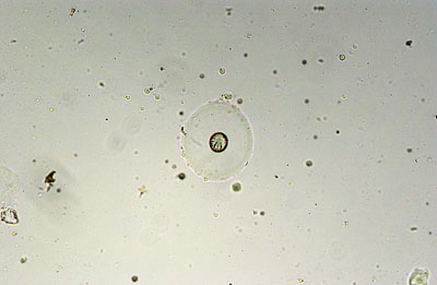
Cestode egg of an iguana
ObjectiveLens 10x
Microscope:Olympus CHS-F DPLAN 10x, Bright-field(Koehler illumination)
Camera:Nikon D1x Lens:NFK2.5x LD
Most of protozoan and nematode eggs are detectable by 10x objective lens. This magnification is often used for screening. Not enough resolution for detail observation.
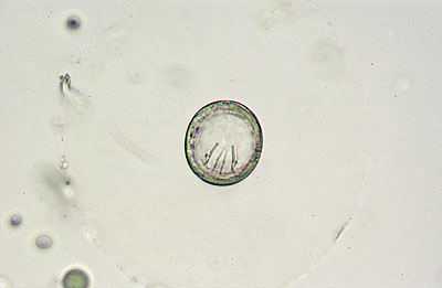
ObjectiveLens 40x
Microscope:Olympus CHS-F A40x PL, Bright-field(Koehler illumination)
Camera:Nikon D1x Lens:NFK2.5x LD
If you use 10x eye-piece, you will have total 400x magnification. By this magnification, more detail and inner structure are able to observe.
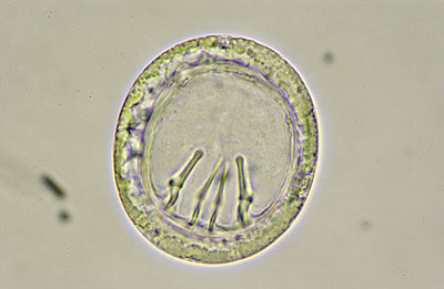
ObjectiveLens 100x
Microscope:Olympus CHS-F DPLAN 100x(Oil), Bright-field(Koehler illumination)
Camera:Nikon D1x Lens:NFK2.5x LD
If you want to observe more details, use 100x(Oil) lens. Close 20-30% of condenser iris to extract maximum performance of your objective lens.
Search for a place where there are less object in the field and set the custom white balance to get the correct white balance. This is the most useful merrit when using digital camera. Do not change the light control after the white balance is corrected. If you have touched the light volume, correct the white balance again.
Difference by condenser iris
Cestode egg of an iguana
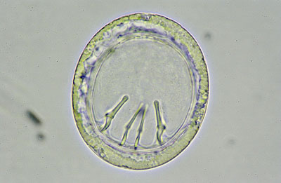
FullOpen
Microscope:Olympus CHS-F DPLAN 100x(Oil), Bright-field(Koehler illumination)
Camera:Nikon D1x Lens:NFK2.5x LD
You can extract full performance by closing iris little bit from full open but depth of field will be very shallow. Strict focus adjustment will be needed.
Smallest
Microscope:Olympus CHS-F DPLAN 100x(Oil), Bright-field(Koehler illumination)
Camera:Nikon D1x Lens:NFK2.5x LD
Depth of field will be deeper but resolution will drop by diffraction.
Difference by illumination-I
Cestode egg of an iguana
BrightField
Microscope:Olympus CHS-F A40x PL, Bright-field(Koehler illumination)
Camera:Nikon D1x Lens:NFK2.5x LD
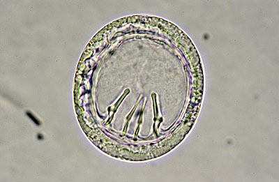
Bright field is the standard illuminating method but it is difficult to observe transparent object. There is a transparent round material around the egg shell above but it is hardly seen by bright field illumination.
PhaseContrast
Microscope:Olympus CHS-F A40x PL, Phase-contrast illumination
Camera:Nikon D1x Lens:NFK2.5x LD
By using phase contrast lens and phase contrast illumination, transparent material can be seen if refractive index is different. Since there is no need to fix or stain, phase contrast method is best to observe live protozoan. This image is same cut with the image above. Round transparent material is clearly seen.
DarkField
Microscope:Olympus CHS-F A40x PL, Dark-field illumination
Camera:Nikon D1x Lens:NFK2.5x LD
As you can enhance light reflecting material by dark field, it is good to use when checking small particle existance. Depth of field will be very shallow as you can not control the iris. Strict focus adjustment is needed.
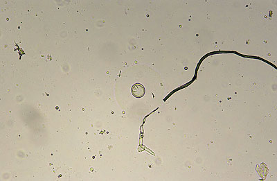
Difference by illumination-II
Cestode egg of an iguana
BrightField Illumination
Microscope:Olympus CHS-F DPLAN 10x, Bright-field(Koehler illumination)
Camera:Nikon D1x Lens:NFK2.5x LD
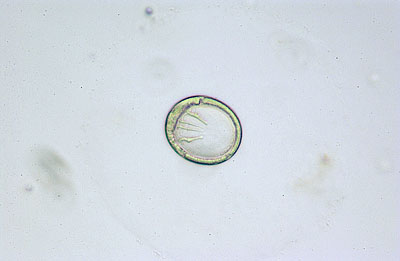
Standard illumination for 10x objective lens. Transparent material is hardly seen.
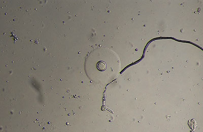
Focal Illumination
Microscope:Olympus CHS-F DPLAN 10x, Bright-field(Focal)
Camera:Nikon D1x Lens:NFK2.5x LD
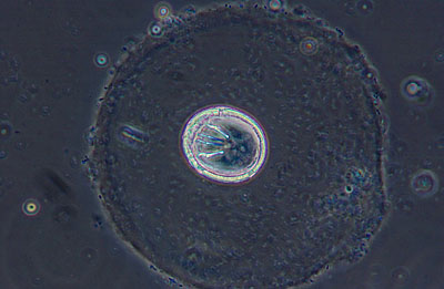
You can get 3-dimentional image by eccentrically placed condenser. There will be a shadow if refractive index of transparent material and the medium is different. Round material can be seen which was not seen in normal bright field.
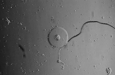
Focal Illumination(Image processed)
Microscope:Olympus CHS-F DPLAN 10x, Bright-field(Focal)
Camera:Nikon D1x Lens:NFK2.5x LD
You can enhance the image by using image processing software such as Photoshop. This image was transformed to gray scale and contrast enhanced.
This is a good way if the lens and/or condenser is poor quality and has chromatic oberration.
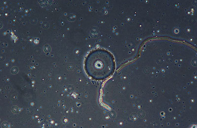
PhaseContrast Illumination
Microscope:Olympus CHS-F A10x PL, Phase-contrast
Camera:Nikon D1x Lens:NFK2.5x LD
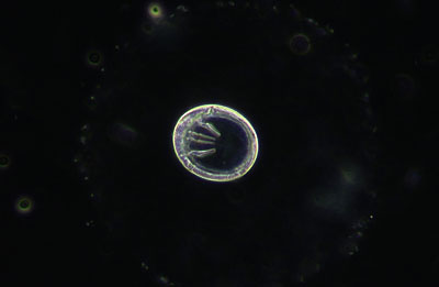
Same cut with phase-contrast illumination. refractive index difference will be transformed to contranst difference. Good to use when observing cilia or flagella of protozoan.
OperationRoom
ImageSample(Ope)
ResolutionTest
Hardware