|
Yamanouchi Iguana Laboratory Video Study for Veterinary Education |
|---|

|
Yamanouchi Iguana Laboratory Video Study for Veterinary Education |
|---|
| Copyrights of all the images(*.jpg, *.wmv) in this page are held by Akira Yamanouchi. If you wish to use the image for educational use, please contact to YIL( yama@yil.jp ). |
| Iguana | ||
|---|---|---|
| Video clip | Title | Comment |
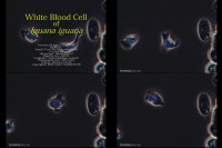 |
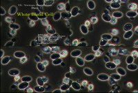
White Blood Cell
Live white blood cell under phase contrast. |
Phase contrast image of live white blood cell. 10x speed. Microscope : Olympus CH-2 Objective Lens : A40PL Camera : Olympus CS230 Place : YIL (KANAGAWA JAPAN) Date : 2006/04/12 Format : 720x480 WMV 1Mbps Size : 7380KB |
 |
White Blood Cell
Live white blood cell under phase contrast. |
Phase contrast image of live white blood cell. 10x speed. Microscope : Olympus CH-2 Objective Lens : A40PL Camera : Olympus CS230 Place : YIL (KANAGAWA JAPAN) Date : 2006/04/12 Format : 720x480 WMV 1Mbps Size : 28647KB |
 |
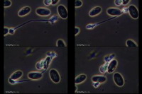
Spermatozoan
Spermatozoan under different illumination. |
Live spermatozoan image. Microscope : Olympus CH-2 Objective Lens : A40PL Camera : Olympus CS230 Place : YIL (KANAGAWA JAPAN) Date : 2006/04/12 Format : 720x480 WMV 1Mbps Size : 28647KB |
| Bird | ||
| Video clip | Title | Comment |
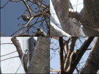 |
Dendrocopos kizuki
Japanese Pygmy Woodpecker. |
Camera : SONY HVR-Z5J Place : Sakai river (KANAGAWA JAPAN) Date : 2009/01/20 Format : 720x540 WMV 1Mbps Size : 9447KB |
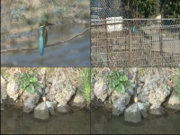 |
Alcedo atthis
Common Kingfisher. |
Camera : SONY HVR-Z5J Place : Sakai river (KANAGAWA JAPAN) Date : 2009/02/06 Format : 720x540 WMV 1Mbps Size : 16570KB |
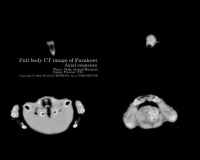 |
CT Image of Parakeet
Axial sequence. |
CT : Toshiba ASTEION Date : 2004/10/20 Format : 400x320 WMV Size : 819KB |
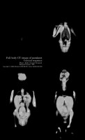 |
CT Image of Parakeet
Coronal sequence. |
CT : Toshiba ASTEION Date : 2004/10/20 Format : 330x540 WMV Size : 1122KB |
 |
CT Image of Parakeet
Sagittal sequence. |
CT : Toshiba ASTEION Date : 2004/10/20 Format : 600x300 WMV Size : 1407KB |
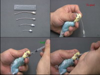 |
Crop Gland Examination
Examination scene on budgerigar. |
Camera : SONY HVR-Z5J Place : EPC (KANAGAWA JAPAN) Date : 2009/01/16 Format : 720x540 WMV 1Mbps Size : 8962KB |
 |
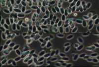
White Blood Cell
Live white blood cell under eccentric illumination. |
Live white blood cell image of budgerigar under eccentric illumination. Microscope : Olympus BX51 Objective Lens : UPLAN40 Camera : Olympus CS230B Place : EPC (KANAGAWA JAPAN) Date : 2009/01/25 Format : 704x480 WMV 1Mbps Size : 9322KB |
 |
White Blood Cell
Live white blood cell under phase-contrast illumination. |
Live white blood cell image of budgerigar under phase-contrast illumination. Microscope : Olympus CH-2 Objective Lens : A40PL Camera : Olympus CS230 Place : YIL (KANAGAWA JAPAN) Date : 2009/01/26 Format : 704x480 WMV 1Mbps Size : 7459KB |
 |
White Blood Cell
Live white blood cell under phase-contrast illumination. |
Live white blood cell image of duck under phase-contrast illumination. Microscope : Olympus CH-2 Objective Lens : A40PL Camera : Olympus CS230 Place : YIL (KANAGAWA JAPAN) Date : 2009/01/29 Format : 704x480 WMV 1Mbps Size : 3396KB |
| Ferret | ||
| Video clip | Title | Comment |
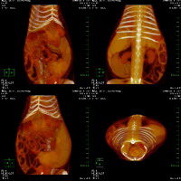 |
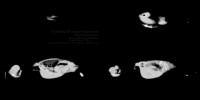
CT Image of Ferret
3-dimentional image of splenomegaly. |
CT : Toshiba ASTEION Date : 2005/04/03 Format : 512x512 WMV Size : 480KB |
| Rabbit | ||
| Video clip | Title | Comment |
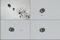 |
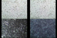
Mange
Mange detected from a rabbit. |
Mange detected from a rabbit. Microscope : Olympus CH-2 Objective Lens : DPlan4 Camera : Olympus CS230 Place : YIL (KANAGAWA JAPAN) Date : 2008/12/13 Format : 704x480 WMV 1Mbps Size : 2802KB |
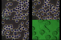 |
White Blood Cell
Live white blood cell under different illumination. |
Phase-contrast image, eccentric illumination image of white blood cell. Microscope : Olympus CH-2 Objective Lens : A40PL Camera : Olympus CS230 Place : YIL (KANAGAWA JAPAN) Date : 2006/05/06 Format : 720x480 WMV 1Mbps Size : 26849KB |